- UGF2,3 Subhash Complex, Tulsidas Marg, opp. Charak Diagnostics Mandi, Chowk, Lucknow, Uttar Pradesh 226003
- Emergency Call: 09794540005 , 09919194111
UGF2,3 Subhash Complex, Tulsidas Marg, opp. Charak Diagnostics Mandi, Chowk, Lucknow, Uttar Pradesh 226003
Emergency Call: 09794540005 , 09919194111
Emergency Call: 09794540005 , 09919194111
Urinary Stone
Urinary Stone Ds
With respect to Division of Stone Disease, we provide treatment for stones in the kidney, the urinary bladder and elsewhere in the urinary tract.
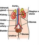
Urinary stones occur in all parts of the urinary system. 97% of all urinary stones are located in the kidney and the ureter. Only 3% are found in the bladder and the urethra.
The following points describe the characteristics of stones:
- Composition : Urinary stones are actually bio-minerals. They contain both inorganic and organic substances. There are different types of stones. Calcium oxalate is by far the most common stone constituent, and is seen in at least 80% of all stones.
- Renal Stones : Renal stone disease is an ancient and common affliction of man. Over a 70-year life-span, it is estimated that about 15% of people will develop renal stones. Although very few individuals die as a direct result of stone disease, it does lead to significant suffering from pain, urinary infections, and obstructive damage to the kidneys. Though newer and more effective methods of stone treatment are available, the actual cause of stone formation frequently remains unknown. So recurrence is bound to take place.
The common symptoms of urinary stone formation are:
- Mild/moderate or severe pain.
- Passage of blood in the urine.
- Passage of tissue in the urine.
- Urinary tract infection.
- Burning sensation when urinating.
- Urinary block.
- No symptoms at all.
Diagnosis of Kidney Stone
Laboratory Tests :
- Complete Blood Count.
- Kidney Function Test.
- Urine Routine & Microscopy.
Ultrasound :
- Whole Abdomen with Kidney, Ureter & Bladder (KUB) .
- X-Ray KUB.
- Intravenous Pyelogram (IVP).
- Plain CT Scan (KUB).
Treatment :
Initial treatment will focus upon the relief of pain. After this, the next step will be to facilitate the passage of the stone or the removal of the stone itself.
80-90% of all stones smaller than 5 mm will pass out on their own. If the stone is smooth, even stones of 7-8 mm may pass out on their own.
Stones larger than this will invariably need to be removed by one of the many methods available. If there is an anatomical abnormality, the priority will be to correct that abnormality while removing the stone.
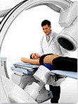
ESWL (Lithotripsy) :
1-2 cm solitary stones in the kidney can preferably be treated by ESWL. uses highly focused electro magnetic waves projected from outside the body to crush kidney stones anywhere in the urinary system. The stone is reduced to sand-like particles that can pass in the urine. Large stones may require more than one sessions. It can be used for patients of all age groups and those who have heart and breathing problems.
PCNL/URS :
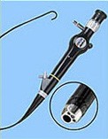
Larger stones in the kidney are preferably removed by PCNL. In this method, the patient needs to be admitted to the hospital. A small puncture is made from the back directly into the kidney, the stone is identified, fragmented and completely removed. Stones lower down in the urinary tract may be treated either by ESWL or again, by endoscopic methods, viz URS. In this, the stone is visualised and fragmented by passing a small endoscope through the urinary opening.
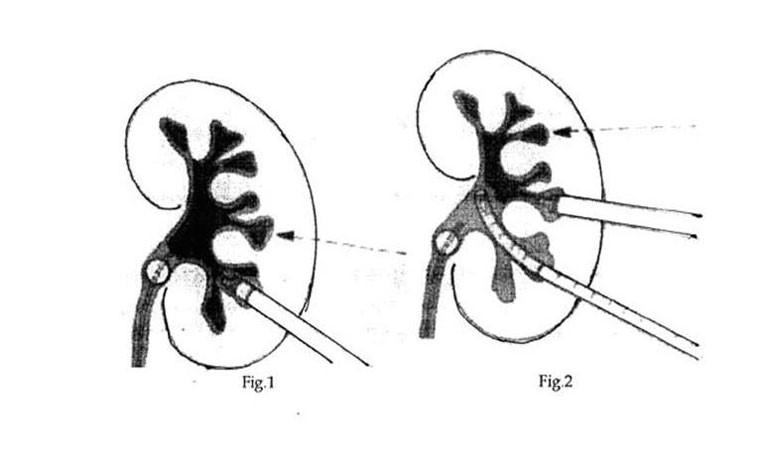
Illustration Showing How PCNL is Performed
Generally, an incision, that is 1 cm or less than 1 cm, is made in the flank. A guide wire is passed through this incision into the kidney. This is performed under fluoroscopy or x-ray control.
A passage is then created around this guide wire by dilatation. Through this passage, a nephroscope is passed into the kidney to visualise the stone and remove it. Larger stones can be fragmented by different methods and removed. Stones are therefore cleared easily. Once the procedure is complete, a tube is left through this tract as drainage for one or two days.
The main advantage of this approach is that, unlike traditional open surgery, only a 1 cm incision is made in the flank. The stones can be visualised directly and removed. The stay in the hospital is only for 3-4 days.
- Ureterorenoscopic Lithotrispy with Holmium Laser is performed under epidural and spinal anaesthesia to treat stones located in the middle or lower ureter. A small, fiberoptic instrument (ureteroscope) is passed into the ureter. Large stones are fragmented using 100-Watt Coherent Holmium Laser. The laser fragments the stone into sand like particles, which are then flushed out through the natural urinary passage. The advantage of Holmium Laser is its ability to fragment stones of all compositions with precision. Thus, it is the most effective laser for the treatment of urinary stones. Patients are generally admitted on the same day of the treatment and are discharged next day, which means only 24 hours of hospitalization is required.
- Prevention of Stone formation : Uric acid stones, which are generally seen only on the ultrasound, and not on the x-rays, if less than 1 cm, can easily be dissolved by simple alkalinisation of the urine.
Most patients with urinary stones need to make certain minimal changes in their diet that may help in the prevention of a recurrence. These include:
- High intake of neutral fluids such as water, tender coconut water, diluted buttermilk, citrus juices, etc., is required unless contraindicated for some other reason. Patients should limit the amount of coffee, tea or milk taken to 1-2 cups a day.
- Food must preferably be vegetarian, and high in fibre.
- Meat eaters should restrict the amount of meat they eat, so that their total protein intake is limited to 1 gram per kilogram per day.
- Cool drinks and soft drinks that contain a high amount of sugar, and calcium-rich foods such as sweets, sweets made using milk as an ingredient, etc., should be avoided, especially on an empty stomach.
Separate and specific changes in the diet may be suggested in the case of other conditions associated with urinary stones.
Patients who are suspected to have other metabolic or endocrine problems will need to undergo detailed testing. These are generally reserved for those patients who have recurrent stone formation.
For Further Information:
- Please go to Urology Health (www.urologyhealth.org) - a patient education site written and reviewed by urology experts in partnership with the American Urological Association Foundation.